ECHOCARDIOGRAMS
Detailed insights of the heart.

Understanding Echos
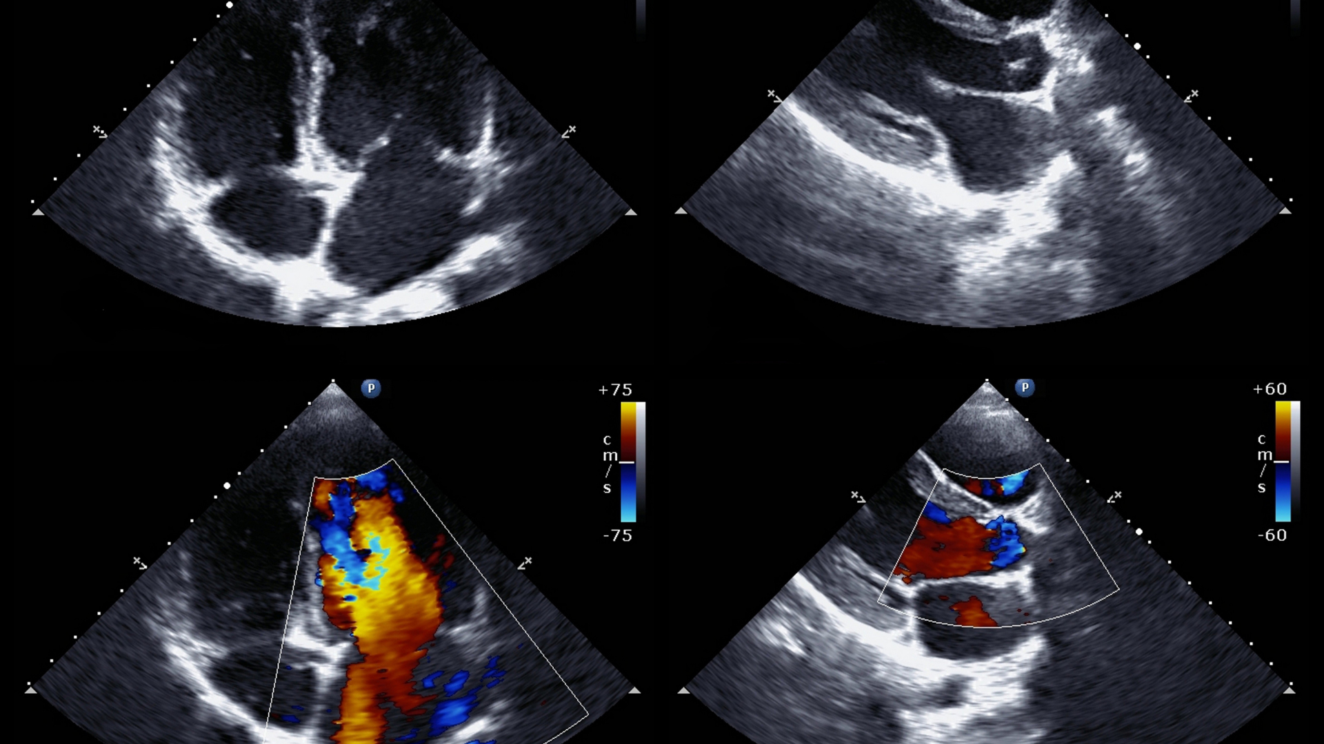
An echocardiogram, often referred to simply as an echo, is a vital diagnostic tool in cardiology. This non-invasive procedure uses ultrasound technology to create detailed images of the heart, allowing physicians to observe the heart’s structure and function in real-time. By emitting high-frequency sound waves, the echocardiogram provides a clear picture of the heart’s chambers, valves, walls, and the blood vessels attached to it. This helps in assessing overall heart health and diagnosing various conditions.
What does it diagnose?
Conditions
Heart Valve Diseases
The echo can detect issues like stenosis (narrowing of valves) or regurgitation (leaking valves), ensuring timely intervention.
Congenital Heart Defects
It identifies structural abnormalities present from birth, facilitating early treatment.
Cardiomyopathy
This procedure helps in evaluating diseases of the heart muscle, including hypertrophic, dilated, and restrictive cardiomyopathy.
Heart Failure
Echocardiograms assess how effectively the heart pumps blood, revealing signs of heart failure and fluid buildup.
Pericardial Diseases
The test can detect inflammation or fluid accumulation in the pericardium, the sac surrounding the heart.
Aortic Aneurysms
It identifies bulges or weaknesses in the aorta, which can be life-threatening if not monitored.
Infective Endocarditis
Echocardiograms detect infections of the heart valves or the lining of the heart, crucial for guiding treatment.

THE PROCEDURE
At Big Sky Imaging, we strive to provide a seamless and reassuring experience for our patients undergoing echocardiograms. Here’s how we ensure exceptional care.
-
Preparation and Consultation:
- Before the procedure, our friendly staff will guide you through the preparation steps, ensuring you are fully informed and comfortable.
- We provide clear instructions on any dietary restrictions or medications to avoid before the test.
-
During the Procedure:
- The echocardiogram is performed by a skilled sonographer who applies a special gel to your chest. This helps the transducer, a device that sends and receives sound waves, to capture clear images.
- The test is painless and typically takes between 30 to 60 minutes. During this time, you will be asked to lie still, and you may be asked to change positions or hold your breath briefly to get different views of your heart.
-
Post-Procedure and Follow-Up:
- Once the procedure is completed, our sonographer and cardiologist will review the images and prepare a detailed report.
- We discuss the results with you promptly, explaining the findings and any necessary next steps in your treatment plan.
- Our team ensures you fully understand your results and provides recommendations for follow-up care if needed.
Why Big Sky Imaging?
Big Sky Imaging is dedicated to delivering high-quality echocardiogram services across Montana. Our state-of-the-art technology, combined with our experienced and compassionate staff, ensures that you receive the best possible care. By providing accurate diagnoses and comprehensive support, we help you manage your heart health effectively.
Find a Location


