An ultrasound, also known as sonography, is a non-invasive imaging technique that uses high-frequency sound waves to produce detailed images of the inside of the body. Unlike X-rays, ultrasounds do not use radiation, making them a safe and widely-used diagnostic tool. The sound waves are transmitted through a transducer, which is a small handheld device that captures the echoes as they bounce off tissues and organs, creating real-time images on a monitor.
Ultrasounds monitor fetal development, assess the health of the baby and mother, and detect any abnormalities.
Abdominal Issues
Used to examine organs such as the liver, kidneys, pancreas, gallbladder, and spleen for conditions like cysts, tumors, or infections.
Cardiovascular Health
Echocardiograms (a type of ultrasound) assess the heart's structure and function, detecting issues like valve problems, heart defects, and cardiomyopathy.
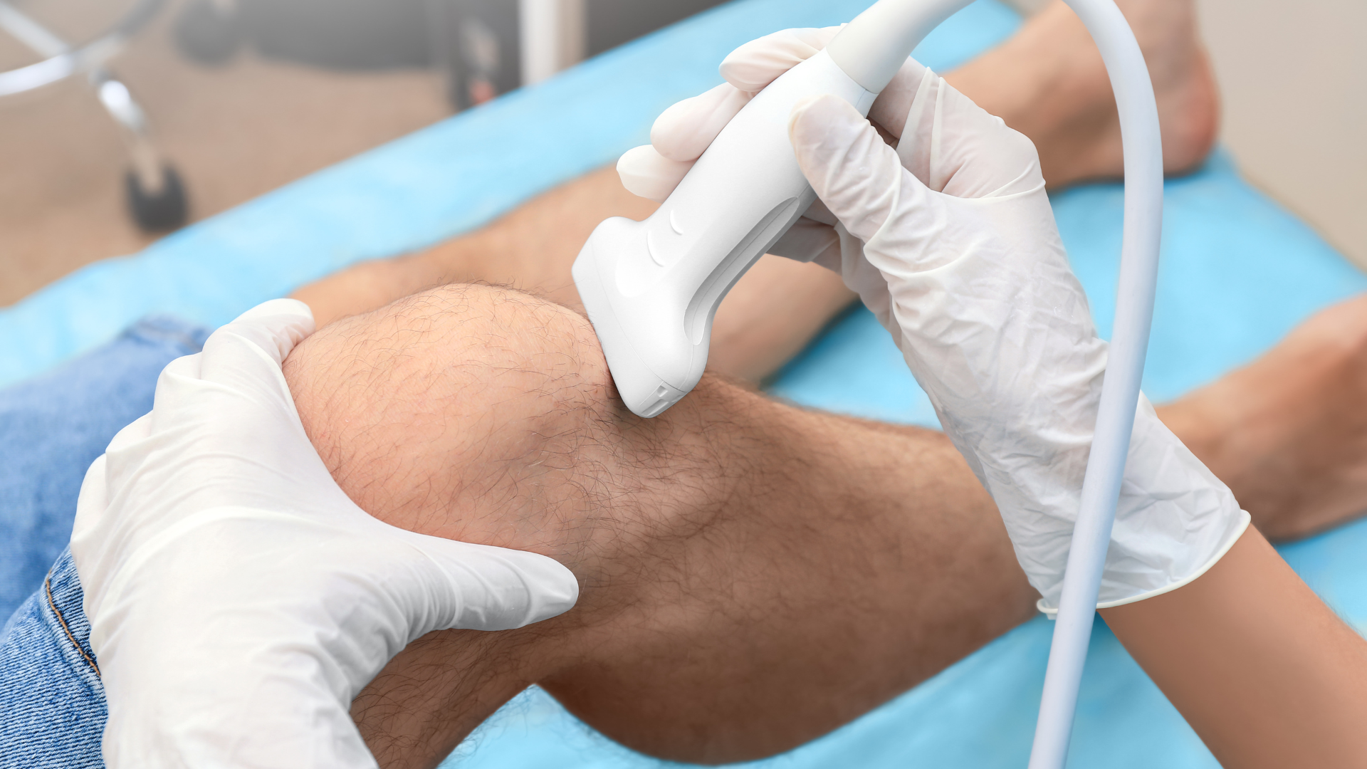
Musculoskeletal Problems
Evaluate muscles, tendons, ligaments, and joints for injuries, inflammation, or other conditions.
Pelvic Health
Examine the reproductive organs in both men and women, detecting conditions such as ovarian cysts, uterine fibroids, or prostate issues.
Vascular Health
Doppler ultrasounds assess blood flow in arteries and veins, identifying blockages, clots, or other vascular conditions.
Thyroid and Parathyroid Glands
Detect abnormalities like nodules, cysts, or tumors in the thyroid and parathyroid glands.
THE PROCEDURE
At Big Sky Imaging, we prioritize patient comfort and accuracy in our ultrasound services. Here’s what you can expect:
Preparation and Consultation:
Our staff will provide you with detailed instructions on how to prepare for your ultrasound, which may include fasting or having a full bladder, depending on the type of exam.
Any questions or concerns you have will be addressed by our team before the procedure.
During the Procedure:
You will be comfortably positioned on an examination table. A special gel will be applied to the area being examined to help the transducer make secure contact with your skin.
The sonographer will move the transducer over the skin to capture images of the targeted area. You may be asked to change positions or hold your breath at times to get clearer images.
The procedure is painless and usually takes between 30 minutes to an hour, depending on the complexity of the examination.
Post-Procedure and Follow-Up:
After the ultrasound, our radiologists will analyze the images and prepare a detailed report.
Results are typically available promptly, and a follow-up appointment will be scheduled to discuss the findings and any necessary next steps.
1 / of3
Why Big Sky Imaging?
Big Sky Imaging is dedicated to delivering high-quality Ultrasound services across Montana. Our state-of-the-art technology, combined with our experienced and compassionate staff, ensures that you receive the best possible care. By providing accurate diagnoses and comprehensive support, we help you manage your heart health effectively.